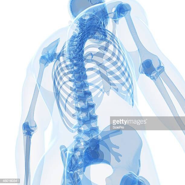
Lumbar Spine

SCIATICA
This is a condition when one of the nerves in the low back (lumbar spine) is compressed. You may experience pain or altered sensation in an area in the leg that is supplied by the involved nerve. In addition you may develop muscle weakness such as difficulty in drawing the big toe or ankle back upwards towards the face. Sometimes this can manifest as a limp. In most cases sciatica settles gradually over time. Only about 10% of patients with sciatica will go on to require surgical intervention.
Causes of Sciatica
Sciatica typically occurs between the ages of 30 and 50.It is typically due to a bulge (prolapse) or rupture of a lumbar disc. Other causes can include compression by a bony spur (osteophyte) or cyst (fluid filled swelling) arising from the facet joints.
Clinical Evaluation
Diagnosis begins with a complete patient history. You will be asked how your pain started, where it travels, and exactly what it feels like.
A physical examination may help pinpoint the irritated nerve root. Tests include assessment of your muscle power by asking you to walk on your heels and toes, and perform a straight-leg raise test.
A magnetic resonance imaging (MRI) scan is the investigation of choice and will help to confirm which nerve roots are affected.
Indications for surgery
Patients who have unrelenting pain that fails to settle are potentially candidates for surgery. In addition patients who present with muscle weakness from the start of their symptoms may benefit from surgery undertaken at a significantly earlier time. This has been shown to improve their chances of muscle recovery.
Treatment options
Physiotherapy
The vast majority of patients with sciatica can be treated simply with pain killers, exercise and physiotherapy. Many patients symptoms begin to resolve quickly within one to two weeks. The majority of patients report a marked improvement in their symptoms over eight to ten weeks from onset.
Spinal injections:
Many patients benefit from x-ray guided (fluoroscopic guided) injections around the involved nerve root to try and decrease pain they are experiencing and decrease inflammation in the nerve root involved. These injections are undertaken as a day case procedure under sedation or local anaesthetic.
Under x-ray guidance a needle is placed close to the nerve and a solution of steroid is injected bathing the nerve and decreasing inflammation around the nerve root. This can take a number of weeks to work and has a success rate of up to 60%. The risks associated with this procedure are low and include infection and nerve injury less than 1/1000 cases.
Surgery
Microdiscectomy
A disc prolapse that compresses a nerve may be dealt with surgically. The standard surgical approach to this is to undertake a microdiscectomy. This is undertaken after the patient is placed under general anaesthesia. The patient is then placed carefully in a prone (face down) position and a small incision is made over the spine at the surgical site. Typically only a small piece of bulging disc is removed allowing decompression of the nerve. Following surgery the wound is typically sutured with a dissolvable stitch and the patient is encouraged to get up and walk as soon as possible.
The patient will typically notice immediate pain relief following surgery if pain was the problem prior to surgery. If however the indication for surgery was weakness this will be slower to settle, often taking weeks or even months for the symptoms to resolve. Post-operative management initially consists of repeated short walks undertaken on an hourly basis, with only gentle physiotherapy undertaken over the first four to six weeks. This is to allow the surgical opening at the back of the disc to heal over before the patient commences formal physiotherapy. Physiotherapy at this stage focuses on building up the back and stomach muscles (core stability program).
Endoscopic discectomy
This is a newer procedure that can be undertaken under sedation using a combination of x-ray guidance and keyhole surgery undertaken using a camera (endoscope). This allows a very tiny incision to be made allowing the patient to recover much more quickly and minimising any trauma to the muscles in the patient’s spine. This decreases the chance of back symptoms after surgery. This is suitable in a specific group of disc prolapse patients.
Post-Operative Management
Following a microdiscectomy surgery you will normally be discharged the following day. You will be allowed to shower ensuring that the waterproof dressing over the wound is intact. However you should not immerse yourself in water e.g bath, Jacuzzi, swimming pool etc until the surgical wound is fully healed which will take between 4 – 6 weeks.
You will be encouraged to walk every hour, gradually increasing your walking distance so that at two weeks after surgery you will typically be able to walk for twenty minutes two to three times per day. Patients can return to driving at two weeks after surgery albeit only for short distances and should avoid lifting weights of more than three to four kilograms for a period of approximately four weeks after surgery. Patients will typically reengage with physiotherapy between four and six weeks after their surgery and return to sporting activities will be dictated by how quickly they progress with physiotherapy.
Back Pain
Soft Tissue Injury
Something as simple as a pulled muscle or strained ligament in the lumbar spine can commonly cause severe pain. This will often come on suddenly (acute onset) potentially following a wide range of activities which can be as minor as bending over.
Thankfully soft tissue injuries can typically be treated with simple measures such as early physiotherapy. This may include measures such as deep tissue massage, dry needling, heat or laser treatments. As the initial early pain begins to settle, a rehabilitation program is recommended to allow full motion of the joints and discs in the spine and strengthen the muscles in the low back that support the spine. In most cases soft tissue injuries will settle within 8 weeks. Symptoms that are not settling within that time frame warrant further evaluation with the investigation of choice being a MRI.

Disc Injury
The intervertebral discs in the spine have a nerve supply of their own (sinovertebral nerve). As we get older the discs in the spine gradually dehydrate and wear out. This can result in tears of the outer envelope of the disc which ca cause severe back pain which may radiate to the leg. Typically initial management will include physiotherapy. If symptoms are failing to settle the patient may benefit from a procedure.
Procedures
These may initially include an epidural injection. If symptoms continue despite epidurals and ongoing physiotherapy the next step would be to consider key hole surgery (Endoscopic discectomy) to attempt to repair the disc envelope. This is a minimally invasive surgery and is normally undertaken under local anaesthetic using endoscopic and radiographic guidance.
A small percentage of patients have symptoms that do not settle and may be candidates for a spinal fusion procedure which again can typically be undertaken using minimally invasive or key hole techniques. This allows very rapid recovery and minimises patient discomfort.
Facet Joint Arthritis
The facet joints are small cartilage lined joints that lie on the back of each vertebra. The cartilage lining these joints and the soft tissue envelope that surrounds them can degenerate and become painful as we age. This can give rise to ongoing back pain, which is classically worse on straightening up from a bent over position.
Procedures
Treatment options for facet arthritis include ongoing physiotherapy. If symptoms are failing to settle with physiotherapy a number of procedures are possible. These initially involve injections of the nerve supplying the joint (rhizolysis) which can offer relief for periods of up to 6 months. This procedure is undertaken as a day case procedure under sedation in the operating room using x ray guidance.
If symptoms fail to settle next option to consider is rhizotomy. This procedure is again undertaken as a day case under sedation using x ray guidance. In this procedure a small electrode is used to ablate the nerve supply to the joints. This blocks pain signals from the nerves supplying the joint offering pain relief for up to 2 – 3 years.

Fractures
Spinal fractures may occur following a severe trauma or a more minor force. The key issues to address are ongoing pain, compression of nerves, any deformity that has occurred as a result of the fracture or the risk of further displacement or movement at the fracture site. Key initial investigations to assess the degree of damage include a MRI and / or CT. Further tests may include an assessment may include an evaluation of the patient’s underlying bone mineral density with a DEXA scan.
Tumours
Tumours are a rare cause of back pain. They may be associated with ongoing severe back pain. This may wake patients at night from sleep. They may also cause neurological symptoms such as pain, altered sensation or weakness in a particular nerve distribution.
Patients may present with other symptoms related to another tumour such as weight loss, altered bowel habit, or palpable mass. Patients with this presentation warrant urgent work up which will typically include a MRI unless this is not possible due for example to an implanted spinal cord stimulator.

Infections
Spinal infections may spread from other areas around the body, travelling in the bloodstream to the spine. The disc spaces are particularly vulnerable due to their limited blood supply. Infection may cause increasing damage to the structures in the spinal column including the discs and bones which can give rise to severe pain. Patients may or may not have obvious signs of sepsis such as raised temperatures, shivering or sweating.
SPINAL STENOSIS
Over 90% of the population over the age of 50 has degenerative changes in the spinal column. As we age the intervertebral discs degenerate and lose their fluid content. This causes them to collapse, bulging backwards into the spinal canal. Compression of the nerve roots can occur due to a combination of a disc bulging from the front and / or spurs of bone which arise from the facet joints at the back of the spine. This can result in the spinal canal becoming narrowed causing compression of the nerve roots. The symptoms the patient experiences depend on the level in the spinal column at which narrowing occurs.

Clinical Evaluation
Patients with spinal stenosis may present with a range of symptoms including:
Back pain: this can arise from degenerative changes in the fact joints or discs in the spine. In addition increased strain on the soft tissues (muscles, ligaments and tendons) can give rise to injury and pain.
Leg Pain: Patients with spinal stenosis are often pain free at rest but note the onset of leg or calf pain with exercise which can limit their walking distance. They can however occasionally use a bicycle or exercise bike without difficulty. Other “tricks” to minimise symptoms can be leaning on a shopping trolley whilst grocery shopping.
Changed sensation such as numbness, burning, or tingling in buttocks, legs or feet.
Weakness in legs or drop foot.
Examination assesses the patients spinal column, neurological system and circulation.
MRI is the initial investigation of choice.
Nonsurgical Treatments
Nonsurgical treatment options focus on spinal rehabilitation and pain management. The underlying narrowing of the spinal canal is unchanged by non-operative methods . However many people report that these treatments do help relieve symptoms.
Physiotherapy: This may comprise strengthening and stretching of lumbar and abdominal muscles, and massage.
Acupuncture: Acupuncture can help reduce muscle spasms and back pain that can accompany spinal stenosis but there is no evidence to demonstrate that it reduces the symptoms of spinal stenosis.
Laser: there is evidence to support the use of laser therapy to address back pain
Anti-inflammatory medications may help reduce swelling around a nerve and reduce pain over a 7 – 10 day course. These medications include Nurofen, Voltarol, and Difene.
Long term use can cause complications including gastritis or stomach ulcers, kidney and cardiac problems. You should check with your doctor if you have a history of cardiac problems, asthma, or gastric problems before starting these medications and stop them immediately if you develop any abdominal pain.

Steroid Injections
Injections of cortisone around an inflamed nerve root can help dexcrease swelling and pain in the nerve. This is typically undertakne in ttheatre under sedation as a day case procedure.
Surgical Treatment
Patients who have not made progress with non operative management and continue to experience day to day difficulty in terms of leg weakness or difficulty walking due to leg pain may be candidates for surgery. The mainprinciple behind surgery is to increase the space available for the nerves and there are a rage of surgical procedures that can achieve this.
Spinal Decompression
Laminectomy (Spinal Decompression). This is a long standing procedure procedure that involves “de-roofing” the spinal canal by removing the lamina. The lamina is removed along with ligaments and osteophytes (bone spurs) that together compress the nerves. This is typically an open surgery and take about an hour to complete.
Spinal fusion
If the spine is unstable prior to surgery or after surgery where extensive bine has had to be removed to decompress the nerves a spinal fusion may be required. This involves inserting screws and rods into the spine to hold the bones in a stable position until they fuse together.
Minimally Invasive Decompression
This technique uses less invasive techniques where a smaller incision is made resulting in less trauma to the tissues and more rapid recovery. The typical hospital stayis two days and patients commence walking n the day of surgery. Magnification is used including the use of microscopes to see the area for surgery.
Interspinous Stabilisation
This technique involves placing a small spacer device between the spinous processes in the back of the spine. These devices hold the vertebrae apart and increase the size of the spinal canal, making more room for the nerves. This procedure is minimally invasive and is often combined with a minimally invasive decompression. In select patient groups success rates are greater than 80%.
Rehabilitation
Following surgery, you may be in the hospital for a number of days depending on your overall health and the procedure you underwent. Walkig id the main activity following spinal decompressive surgery with physiotherapy commencing 4 – 6 weeks after surgery.
This is focussed on developing strength and flexibility in the muscles of the back and abdomen.

CAUDA EQUINA SYNDROME
Cauda Equina Syndrome occurs when a disc in the spine bulges backward causing severe compression of the nerves below the L1/2 level. The exact nature of the patient symptoms depend on the level at which the compression has occurred but will frequently include 3 cardinal symptoms:
Bilateral Sciatica (pain down both legs)
Saddle anaesthesia (altered sensation in the private parts)
Loss of urinary continence
